
Taking A Closer Look At Your Eyes and Vision
Your vision – the act of seeing – is incredibly complex. In fact, your eyes are on their “A” game 24/7. Yet, even those who need glasses or contacts to see well often take their eyes and vision for granted. The eyes just…make it happen. The reality is what “happens” is amazing! And, for those weighing their vision correction options, understanding how the eye works can be helpful in making an informed decision.
Eye Doctors
Many people with vision problems work with an eye doctor for routine vision check-up and glass prescriptions. Many patients don’t realize there are several types of ophthalmologists – medical doctors and eye surgeons – who specialize in different parts and functions of the eye and vision. Often, when significant changes or challenges occur, patients will be referred to a physician or surgeon with a medical degree to help protect and improve the health and function of the eye. Eye exams performed by ophthalmologists evaluate the entire eye and visual system with a variety of sophisticated diagnostic instruments that go beyond what patients typically experience in a routine vision check-up.
This is particularly true for those patients seeking to move beyond glasses and contacts to a more permanent solution to their vision problems with a laser vision correction procedure such as LASIK. Refractive surgeons – highly trained ophthalmologists – are qualified to perform laser vision correction procedures. These procedures happen in the very front of the eye, or cornea, where the process of vision begins.
How Do Your Eyes and Vision Work?
How does vision work? First, light enters through the cornea at the front of the eye. The cornea is responsible for the majority of the eye’s focusing ability. Then, light is then filtered and directed to the crystalline lens which sits just behind the pupil. The lens sharpens the focus of the light as it travels to the retina at the back of the eye. The retina converts the light rays – the image – into electric signals that are picked up by the optic nerve. Finally, the optic nerve sends those electric pulses to the part of the brain that controls our sense of vision, the visual cortex, to complete the process of seeing.
Because laser vision correction is an option many people consider, it is helpful to explain the anatomy at the front of the eye where these procedures take place.
The Cornea
The clear surface of the front of the eye is known as the cornea. It functions like the crystal on a watch, curving around the front of the eye to focus light like a camera lens. The cornea bends—or refracts— the light onto the crystalline lens. In fact, the cornea accounts for 65 – 75 percent of the eye’s total focusing power. This is why most laser vision correction procedures occur within the cornea. Nearsighted people have a cornea that is curved too steeply, meaning the distance between the cornea and the retina is too long. Nearsightedness – a.k.a. myopia affects about 30 percent of the population. Approximately 10 percent of people in the U.S. are farsighted. Also known as hyperopia, farsighted people have corneas that are too flat, meaning the distance between the cornea and the retina is too short. The cornea can also be curved irregularly, which causes astigmatism. People with astigmatism may also be nearsighted or farsighted.
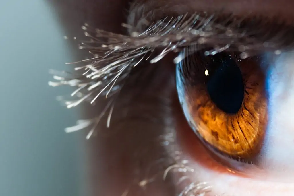
The Iris and Pupil
The iris and the pupil work together to control the amount of light that comes into the eye. The iris is the colored part of the eye that surrounds the dark pupil at the center. The iris adjusts the size of the pupil in response to the amount of light coming through the cornea.
The Crystalline Lens
Located directly behind the pupil, the lens of the eye further focuses light. This small, clear, disc-shaped structure changes its shape to focus on images at different distances. It works like an autofocus camera lens that moves to bring objects into focus. With age, the lens loses its flexibility, making it difficult to see things up close. This is why most people need reading glasses after the age of 40 (and not because a vision correction procedure wore off). It is a condition called presbyopia – greek for “aging eyes.”. The lens is also the part of the eye where cataracts develop. The lens becomes cloudy, making it difficult to see clearly. These age-related changes to the lens are why most people in their 40s and beyond need vision correction for reading.
Options for Your Eyes and Vision
Refractive surgery is just one of many specialized fields in ophthalmology. Refractive surgeons dedicate their practice to restoring the eye’s focusing ability to improve vision. While laser vision correction procedures like LASIK, SMILE, and PRK, are the most well-known and performed, refractive surgeons are trained in many technologies and techniques, such as implantable collamer lenses (ICL), cataract surgery, corneal inlays, and more, that treat the parts of the eye that affect focus and vision.
Other specialties in ophthalmology include general, pediatric, retina, as well as diseases such as glaucoma and macular degeneration. These highly trained medical doctors commit years, if not decades, to honing their skills and expertise to help patients protect the health of their eyes and improve their vision.
Working with an ophthalmologist and having a thorough annual eye exam is important, whatever eye or vision issue a patient may experience. If you are thinking about your laser vision correction options, read to learn what to look for in a refractive surgeon.